THE SURGERIES START at 7:30. Anxious patients are wheeled into more than 20 operating rooms. The anesthesia kicks in. Scrub nurses hand surgeons their scalpels and the first incisions are made. Soon after that, they begin to fill the plastic containers – some as small as jelly jars, some as big as buckets. In goes a gallbladder. A uterus. The head of a femur. Part of a stomach. A testicle. Half a thyroid. A colon. A liver. The “specimens,” as these excised body part are now known, are then delivered to the nearby “gross room,” which is overseen by the department of pathology.
As a third-year resident in pathology, I’ve spent countless hours over the past few years in the gross room. It’s a far cry from the newsrooms I used to work in – I was a journalist for more than a decade before going back to school and earning a medical degree – but increasingly I’m learning the ropes.
Given what we do here, most non-medical people would consider “gross room” an apt description. But the word “gross” is used here the same way it is in “gross anatomy,” meaning that which is visible to the naked eye, without the aid of microscopes.
Pathologists specialize in the diagnosis of disease, using a variety of methods from scrutinizing cells under a microscope to state-of-the-art analysis of a patient’s genetic makeup in a search for chromosomal abnormalities. This essential but largely behind-the-scenes role has led some to describe them as the most important doctors you’ll never meet.
Evaluating specimens removed in surgery to determine what afflicts them and to what degree begins in the gross room. It is a crucial part of a pathology resident’s training, done under the tutelage of attending physicians and pathology assistants who know these organs like the back of their hands.
The process is known as, yes, “grossing.”
~
It’s something patients may not think about much when they go into surgery—exactly what happens to body parts after they’re removed.
At my hospital, the grossing is done at one of five grossing stations which are equipped with a cutting board and a set of tools including scalpels, forceps, sharp scissors, scales and rulers. We wear gloves and plastic gowns over our scrubs as we analyze the specimens and dictate our descriptions into a “gross report,” using words such as “necrotic” (dead), “purulent” (filled with pus), “friable” (crumbly) and “hyperemic” (bright red, as if filled with blood) that shed insight into the patient’s condition.
The long hours of medical residency training are intended, in part, to maximize exposure to patients so physicians can be prepared for almost anything that may come their way in practice. For obstetricians-in-training that means delivering innumerable babies. The experience can be at times repetitious, but through each experience you gain a more nuanced understanding of the process. And you never know when the unexpected will occur.
For future surgical pathologists, it’s grossing multitudinous specimens and, later, examining them under a microscope. The goal for any doctor, one clinician explained to me during medical school, is that over time doing the right thing becomes so instinctive that you practically could do it in your sleep – but the principle that constant repetition leads to proficiency can be applied to anything from learning to play violin to perfecting a jump shot.
In the gross room, the scope of the specimens grossed each day can be staggering – from routine infected appendices to pancreatic cancers removed along with portions of adjacent small intestine and stomach from a complicated surgery known as a “pancreaticoduodenectomy” (aka a “Whipple procedure”).
~
It’s not all cancer in the gross room. On any given day, you may see a thyroid gland with multinodular goiter, a uterus with massive fibroids, a cirrhotic liver, lungs from a cystic fibrosis patient undergoing a transplant, a foot that had to be amputated because of frostbite or a small intestine riddled with bullet holes from Chicago’s inner-city gang warfare.
But cancer is a big part of it. And after doing this for almost three years, I’ve learned what cancer looks like.
Sometimes, it’s a papillary, cauliflower-like growth inside a bladder. Sometimes it’s a fungating tumor in the colon, like an ugly mushroom blocking the flow. Sometimes it’s a dark, irregularly-shaped melanoma spreading just beneath the surface of the skin.
There are many different cell lines in the body and different cancers affect the body in different ways. Some are far more aggressive than others and some are harder to treat and contain.
But all cancers share something in common: one kind of cell in the body has gone haywire and is spreading to the point where it edges out the normal cells that are just trying to do their job. Body functions are compromised and the consequences can be grim.
There’s a lot of cancer to take in and process in our job. You can’t help thinking about mortality and the cruel fates of who gets it and why. Lymphoma in a 27-year-old grad student. Ovarian cancer in a 38-year-old mother of three. It could easily make our work morose – after all, cancer is the second-leading cause of death in the United States, trailing only heart disease.
If there’s a mitigating factor, it’s that we in pathology generally do not meet our patients, so we always bear in mind that behind that thyroid, or that uterus, or that bladder, is a nervous patient trusting that we will treat the specimen right and come up with the proper diagnosis.
~
I was thinking about Walter White as I sliced into a lung and revealed a firm, white nodule standing out against the normal pink spongy inside of a lung. It was, unmistakably, cancer. At the beginning of “Breaking Bad,” the high school chemistry teacher learns that he has Stage IIIA lung cancer. That ominous diagnosis irrevocably rocks his world and sets the stage for radical decisions that dramatically alter the trajectory of his life.
Walter White is a fictional character, but the right upper lobe of lung I am examining is all too real. So too is the forthcoming diagnosis, which will change a real patient’s life, as hearing a doctor speak the word “cancer” inevitably does.
At the end of the process I am beginning, the patient will learn the stage of his cancer, a determination which will set the course of his treatment and provide him with a prognosis. In other words, he’ll find out what his odds are of beating the disease, and for about how long.
Often, when an operation occurs, the patient already knows he has cancer because of an earlier biopsy. So part of our job in the gross room is to find out how far it’s gotten. To document this, we take careful standardized steps.
For instance, when we receive a lumpectomy—an excision of breast tissue containing a tumor—we begin by applying brightly colored dyes to each side of the specimen, representing the points at which the surgeon made the cuts. Red, orange, yellow, green, blue and black—painted directly onto the fatty breast tissue.
So, for a few moments, our job is kind of like an odd little arts and crafts project. But the subject matter is serious. Not much in life is more serious than cancer.
The point of this is when we cut into the specimen, we will remove small portions of the tissue—each about the size of a postage stamp—which will be placed into a small plastic cassette, fixed in paraffin wax and thinly sliced to make slides.
Attending pathologists looking at those slides under a microscope later will make the definitive diagnosis of the exact kind of cancer. But also, because of the colorful dyes, pathologists can tell if the tumor is a safe distance from those surgical margins. This is crucial. It’s the determination that allows the surgeon to tell the patient, “Yes, it’s cancer, but we believe we removed it all.”
~
How far has the cancer gotten? The staging of the cancer is a succinct summary of where the patient stands—which pathologists determine after the grossing and the microscopic analysis of slides. To determine that, we use the “TNM” staging system.
T is for tumor—how big is it? Does it invade the organs next door?
N is for lymph nodes. Have the cancer cells spread into lymph nodes near or far?
Pathology residents are often tasked with determining this by sorting through the specimen to look for lymph nodes. In some parts of the body, they’re as big as lima beans so they pop right out at you. In other areas, it’s the anatomical equivalent of finding a needle in a haystack.
Many rectal cancer patients are treated before surgery with radiation that shrinks their lymph nodes to the size of poppy seeds. What’s more, they’re buried within mounds of “adipose tissue” – basically, we’re talking fat—attached to the colon as it dips into the rectum. The nodes blend in with the surrounding tissue, but have a slightly different glisten and a somewhat firmer consistency.
Colorectal cancer studies have determined that you need to pluck out at least 12 lymph nodes to provide a big enough sampling to tell whether the cancer has spread there – and if it has, the subsequent treatment of the patient is stepped up. Residents develop their own techniques to find nodes. Some use scalpels to cut razor-thin slivers through every centimeter of fat and keep their eyes peeled. Others use their gloved fingers to squeeze every inch of the fat to see if they can detect something that is just a tad more solid.
Being a doctor is a very glamorous job.
The M stands for “metastasis.” Has the tumor turned up in other organs farther afield—for instance, has the colon cancer spread to the liver? The treatment is more complicated, and the prognosis less optimistic, if the cow already has gotten out of the barn.
~
When the grossing is finished, we place the specimen back into its container and soak it in formaldehyde to preserve it in case we need to pull it out and re-examine it later. Those little samples of tissue we took from it are on their way to becoming slides. The next steps in diagnosing the disease will occur under the microscope.
Tomorrow morning, the surgeons will be busy again and the specimens will begin to trickle in. A prostate. A breast. An ovary. A kidney. A toe.
We’ll be ready in the gross room.
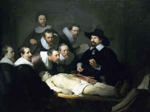
Rembrandt, The Anatomy Lesson of Dr. Nicolaes Tulp, 1632